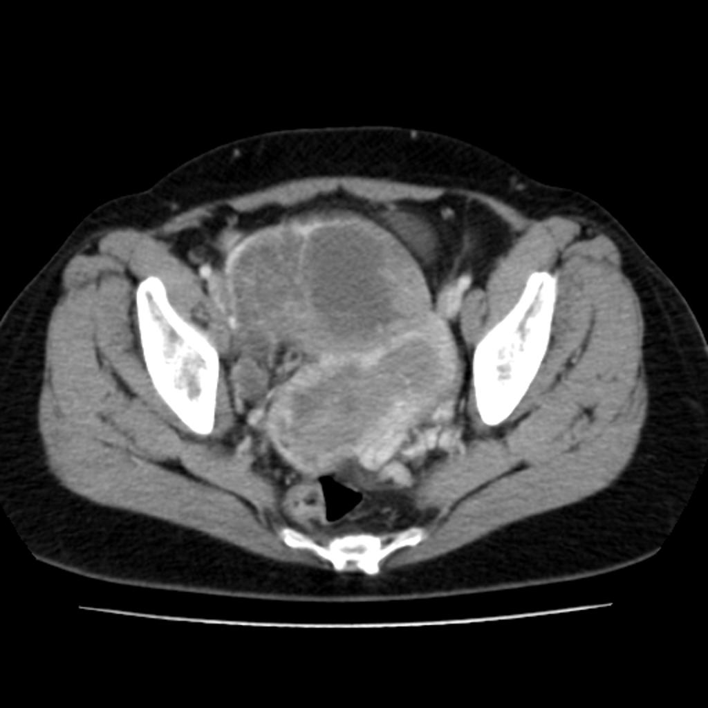
| Stade I | Limité au corps utérin et à l’ovaire
|
| IA | Disease limited to the endometrium OR non-aggressive histological type, i.e. low-grade endometroid, with invasion of less than half of myometrium with no or focal lymphovascular space involvement (LVSI) OR good prognosis disease |
| IA1 Non-aggressive histological type limited to an endometrial polyp OR confined to the endometrium |
| IA2 Non-aggressive histological types involving less than half of the myometrium with no or focal LVSI |
| IA3 Low-grade endometrioid carcinomas limited to the uterus and ovary |
| IB | Non-aggressive histological types with invasion of half or more of the myometrium, and with no or focal LVSI |
| IC | Aggressive histological types limited to a polyp or confined to the endometrium |
| Stade II | Invasion of cervical stroma without extrauterine extension OR with substantial LVSI OR aggressive histological types with myometrial invasion |
| IIA | Invasion of the cervical stroma of non-aggressive histological types |
| IIB | Substantial LVSI of non-aggressive histological types |
| IIC | Aggressive histological types with any myometrial involvement |
| Stade III | Local and/or regional spread of the tumor of any histological subtype |
| IIIA | Invasion of uterine serosa, adnexa, or both by direct extension or metastasis |
| IIIA1 Spread to ovary or fallopian tube (except when meeting stage IA3 criteria) IIIA2 Involvement of uterine subserosa or spread through the uterine serosa |
| IIIB | Metastasis or direct spread to the vagina and/or to the parametria or pelvic peritoneum |
| IIIB1 Metastasis or direct spread to the vagina and/or the parametriaIIIB2 Metastasis to the pelvic peritoneum |
| IIIC | Metastasis to the pelvic or para-aortic lymph nodes or both |
| IIIC1 Metastasis to the pelvic lymph nodesIIIC1i MicrometastasisIIIC1ii MacrometastasisIIIC2 Metastasis to para-aortic lymph nodes up to the renal vessels, with or without metastasis to the pelvic lymph nodesIIIC2i MicrometastasisIIIC2ii Macrometastasis |
| Stade IV | Spread to the bladder mucosa and/or intestinal mucosa and/or distance metastasis |
| IVA | Invasion of the bladder mucosa and/or the intestinal/bowel mucosa |
| IVB | Abdominal peritoneal metastasis beyond the pelvis |
| IVC | Distant metastasis, including metastasis to any extra- or intra-abdominal lymph nodes above the renal vessels, lungs, liver, brain, or bone |
